A PET-CT is recommended when doctors need to know not only where an abnormality is, but whether it is biologically active and how far disease may have spread. By the end, you will understand what the scan can reveal, why it is chosen over other tests, how to prepare, and how its findings can change treatment decisions.
Key takeaways
- PET-CT combines metabolic activity with precise anatomical location.
- A bright PET area does not prove cancer; infection can also increase uptake.
- Follow fasting and blood-sugar instructions before an FDG PET-CT.
- Biopsy results can confirm findings before treatment begins.
What a PET-CT Shows Beyond a Standard Scan
A PET-CT shows both what tissue is doing and where that tissue sits in the body. For a common PET scan, you receive fluorodeoxyglucose, a glucose-like radioactive substance.
The FDG tracer collects more in cells using glucose rapidly; PET detects this signal, while CT supplies detailed anatomy and aligns the uptake with an organ, lymph node, or bone.
| Scan | Main strength | What PET-CT adds |
|---|---|---|
| Diagnostic CT | Shows anatomy, size, and shape | Shows whether tissue is metabolically active |
| MRI | Provides detailed soft-tissue contrast | Maps metabolic activity across the scanned body region |
| Ultrasound | Displays reachable structures in real time | Shows tracer uptake beyond the ultrasound field |
| Bone scan | Highlights abnormal bone activity | Shows metabolic activity in soft tissues and provides CT localisation |
This combined information can reveal active disease before a clear structural change appears, or show that a suspicious area is metabolically quiet. PET-CT can also help identify active tumour for radiotherapy planning, although CT or MRI remains essential for precise boundaries.
Increased uptake is not a cancer diagnosis. Infection, inflammation, healing tissue, active muscles, and some benign tumours can absorb FDG, while slow-growing or low-metabolism cancers may appear less conspicuous. Urinary tracer activity can obscure nearby structures, so doctors interpret PET-CT with clinical findings and other imaging; a biopsy may still be needed.
The Cancer Questions a PET-CT Can Answer
An oncologist may recommend a PET-CT to resolve a decision that anatomy alone cannot settle. The questions often concern the extent or activity of disease:
- Is an indeterminate lesion active disease, or could it be scar tissue or another benign finding?
- Does cancer staging show involvement beyond the suspected primary tumour?
- Do nearby lymph nodes contain active tumour, even if they are not enlarged?
- Is there distant spread to organs or bones that would change local treatment?
- Where is the primary tumour if a biopsy has found cancer but routine imaging has not located its origin?
- Does a residual mass after treatment contain viable tumour, and can the scan establish a baseline for later comparison?
- Has treatment reduced metabolic activity, or does new uptake suggest progression or recurrence?
The result can show where metabolically active tissue lies and help identify a safer, more informative biopsy site. It does not prove that every bright area is cancer: a non-cancerous process can cause uptake, while some slow-growing tumours show little uptake.
The chosen tracer also matters; PSMA-PET, not FDG-PET, is used in selected prostate-cancer staging or recurrence assessments. A recommendation reflects the specific cancer and the unanswered clinical question, not a routine step for every abnormality.
When PET Findings Need Confirmation or a Different Test
Increased FDG uptake is not proof of cancer. Infection, inflammation, healing tissue, active muscles, brown fat, some benign tumours, and urinary tracer activity can all create bright areas or hide nearby lesions. A false positive PET scan can follow recent surgery, radiotherapy, or biopsy because healing tissue remains metabolically active.
SUV provides a measure of tracer concentration, but no single SUV value separates cancer from inflammation in every organ. A scan performed too soon after treatment can also look worse because treatment-related inflammation remains FDG-avid.
The correct interval depends on the cancer and treatment; head-and-neck chemoradiotherapy response scans are commonly timed at about 12 weeks, not immediately after treatment.
Ask what follow-up fits when:
- Prostate cancer is suspected: FDG-PET is often less useful for typical prostate cancer because uptake can be low. A PSMA-PET, which uses a different tracer, may be more appropriate for selected staging or recurrence questions. Do not treat FDG-PET and PSMA-PET results as interchangeable.
- An unexpected focus appears: targeted diagnostic CT or MRI, interval imaging, or endoscopy can show whether anatomy matches the PET finding.
- Treatment would change based on one suspicious spot: a biopsy after PET scan may be needed to confirm viable tumour and establish pathology before surgery, radiotherapy, or systemic treatment.
- The lesion remains unclear: discuss whether respiratory motion, imperfect PET-CT image registration, low spatial resolution, or nearby urinary activity affected interpretation. PET is evidence to interpret with symptoms, examination, laboratory results, and other imaging—not a diagnosis by itself.
How to Prepare for the Appointment and What Happens During the Scan
PET scan preparation usually includes avoiding calories for 4–6 hours before arrival. Drink plain water, avoid strenuous exercise that day, and follow instructions about regular medicines.
Diabetes and PET scan preparation need individual instructions because high blood glucose or insulin levels can reduce image contrast; the centre may change or postpone the appointment if your glucose is substantially elevated. Tell the team about pregnancy, possible pregnancy, or breastfeeding.
1. At check-in, staff confirm your medical history and measure blood glucose. Wear warm, comfortable clothing and remove metal items before imaging.
2. The PET scan injection places a small amount of radiotracer, commonly FDG, into a vein. Afterward, rest quietly for roughly 45–60 minutes; talking, walking, and muscle activity can affect tracer distribution.
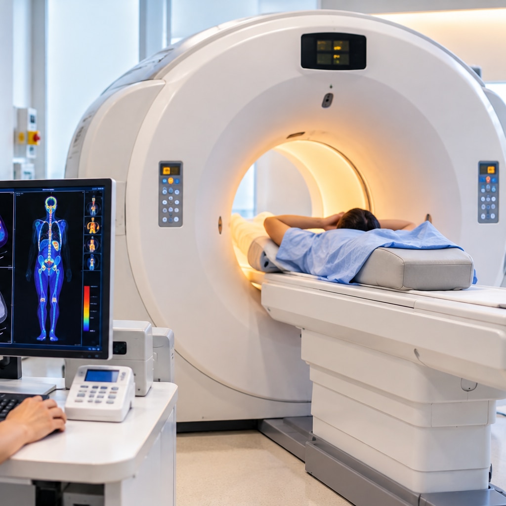
3. You lie on your back on a narrow, padded table. The CT portion usually comes first, followed by the PET images, as the table moves slowly through the scanner. Keep still and breathe normally unless staff give another instruction.
4. Imaging often takes 20–30 minutes. Tell the technologist if you feel unwell, become uncomfortable, or cannot remain still.
The CT may be a low-dose scan for mapping the tracer or a diagnostic scan with different radiation exposure. Afterward, drink water and urinate regularly unless your clinical team gives different advice.
How PET Results Can Change the Treatment Plan
A PET-CT can move the plan from treating one visible tumour to treating disease throughout the body. If it shows metabolically active lymph nodes or distant deposits, surgery or curative radiotherapy aimed only at the original site may no longer address the full disease burden; systemic treatment may become the priority.
It can also identify the safest, most informative site for biopsy.
- Confirm what the scan changes before acting. Compare the uptake with CT or MRI findings and the clinical history; FDG uptake from infection, healing, or inflammation is not proof of cancer.
- For surgery, ask whether every active focus can be removed with a meaningful benefit. Unexpected spread can prevent an operation that would leave untreated disease, while a clearly localised pattern can support surgery.
- For radiotherapy, a radiotherapy planning PET-CT can help define metabolically active tumour that is difficult to distinguish on CT. The radiation team still checks MRI or diagnostic CT, breathing movement, image registration, and inflammation before setting the treatment volume.
- For systemic treatment, the scan can establish a baseline and later show treatment response. Do not schedule response imaging too soon: post-treatment inflammation can look active, and the correct interval depends on the cancer and therapy.
- An unexpected focus may lead to targeted imaging, repeat imaging, endoscopy, or biopsy rather than an immediate treatment change. During cancer surgery consultation at Dr Gawande Oncocare Centre, PET findings can help test whether an operation fits the disease pattern; later scans may also investigate suspected cancer recurrence.
Frequently asked questions
What does a PET-CT show beyond a standard scan?
A PET-CT shows how actively tissues use glucose and where those tissues are located, combining metabolic and anatomical information.
Can a PET-CT confirm cancer by itself?
No. Infection, inflammation, and other conditions can cause increased FDG uptake, so doctors may need a biopsy or another test.
How should you prepare for an FDG PET-CT?
Follow the centre’s fasting instructions, discuss diabetes medicines in advance, avoid strenuous exercise before the scan, and wear comfortable clothing.
How can PET-CT results change cancer treatment?
Results can help define the cancer’s extent, identify suitable treatment targets, assess response, and show whether surgery, radiation, systemic therapy, or further testing is appropriate.