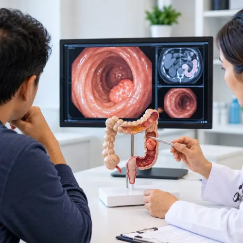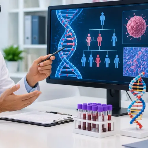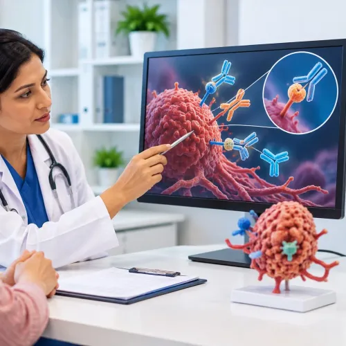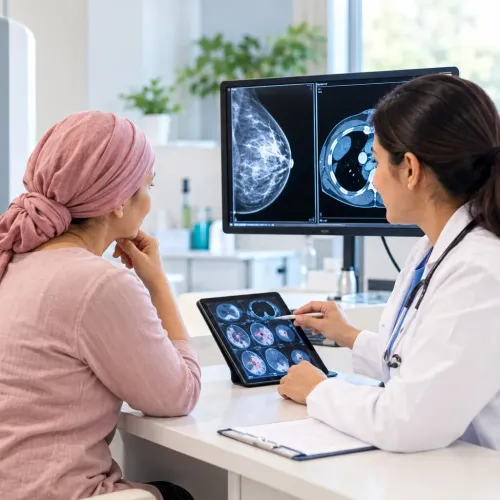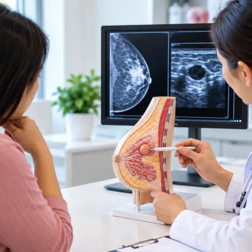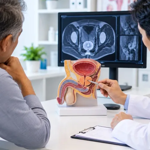
A scan can reveal a lump, thickening, or area that looks abnormal, but it usually cannot identify exactly what that tissue is. By understanding how biopsies are collected, examined, and reported, you can ask informed questions about whether the sample is adequate and what the result means for the next decision.
Key takeaways
- A suspicious scan needs tissue testing before cancer is confirmed.
- The biopsy method depends on the lesion’s location, size, and accessibility.
- Ask when preliminary and final pathology results will be available.
- Read the pathology report for cancer type, grade, and tested biomarkers.
Why a scan cannot usually confirm cancer
A scan cannot usually confirm cancer because images show structure or activity, not the identity of the cells. A suspicious lesion is a finding that needs explanation, not proof of malignancy.
- CT shows size, shape, density, and spread through nearby or distant areas.
- MRI shows soft-tissue detail and how a lesion relates to surrounding structures.
- Ultrasound shows internal features, blood flow, and whether a lump is solid or fluid-filled.
- Mammography shows patterns such as masses, distortion, or calcifications in breast tissue.
- PET shows increased tracer uptake, which can occur with cancer, infection, or inflammation.
A biopsy removes cells or tissue for a pathologist to examine. It can establish whether the sampled area is malignant, benign, infected, inflamed, scarred, or affected by another non-cancerous condition.
That distinction changes the cancer diagnosis: treating an infection or scar as cancer would expose you to unnecessary treatment, while dismissing cancer as inflammation could delay care.
A tissue-confirmed diagnosis can also identify the cancer type and provide material for grade, molecular changes, or biomarkers that guide treatment. The sample must contain enough suitable tissue; a small cell sample can be insufficient when the diagnosis requires tissue architecture.
A liquid biopsy examines tumour-derived material, such as circulating tumour DNA, in blood. It can help when tissue is difficult to obtain, but a negative result does not reliably rule out cancer or replace tissue biopsy when sampling is safe.
Which biopsy method is used and why
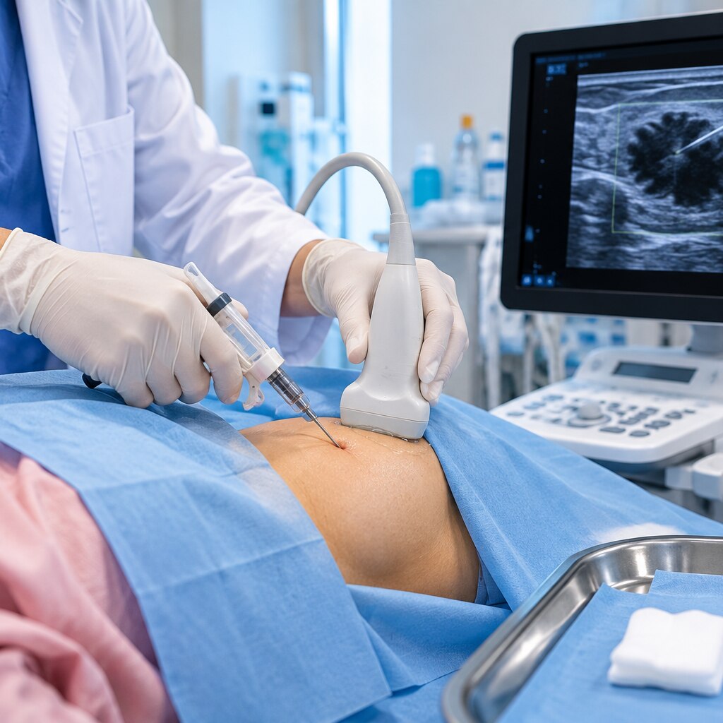
The method depends on the lesion’s location, size, accessibility, and the amount of tissue needed for testing. A fine-needle sample may show abnormal cells, while some diagnoses require preserved tissue architecture from a larger fragment.
| Method | What the sample can reveal | Why doctors choose it |
|---|---|---|
| Needle biopsy | Cells or tissue from a lump or deeper lesion | Used when the target can be reached through the skin with imaging guidance |
| Fine-needle aspiration | Individual cells, fluid, and signs of infection or malignancy | Useful for accessible lumps, cysts, thyroid nodules, or lymph nodes; it may not preserve tissue structure |
| Core needle biopsy | Several tissue cylinders showing architecture, tumour type, grade, and some biomarkers | Preferred when more tissue is needed without removing the whole lesion |
| Endoscopic biopsy | Mucosal tissue from the digestive, respiratory, urinary, or reproductive tract | Chosen when a camera can reach an internal abnormal area |
| Skin biopsy | Surface cells from a shave biopsy or deeper layers from a punch biopsy | Used for suspicious moles, rashes, or skin growths; depth can help assess invasion |
| Surgical or excisional biopsy | A larger specimen, sometimes the entire lesion, with possible margins and invasion assessment | Used when needle access is unsafe, tissue is inadequate, or complete removal is practical |
A negative result does not always exclude cancer. The needle can miss the tumour or collect too little tissue, leading to repeat sampling or a different technique when imaging or symptoms remain concerning.
The expected benefit must be weighed against site-specific bleeding, infection, pain, or injury to nearby structures. A biopsy identifies the sampled lesion; it does not, by itself, establish the cancer’s full stage.
What happens before, during, and after the procedure
1. Before the procedure, consent should explain why the sample is needed, which area will be removed, what the result may and may not establish, and the alternatives. Ask about bleeding, infection, pain, scarring, injury to nearby structures, and the rare possibility of tumour-cell spread along the biopsy path.
2. Anaesthesia depends on depth and location. A local anaesthetic numbs the skin for a punch biopsy, shave biopsy, or many needle biopsies; sedation or general anaesthesia may be used for a deeper surgical biopsy or excisional biopsy. You may feel pressure or pulling, but sharp pain should be reported.
3. The team should confirm your identity, the exact body site and side, and the planned procedure before sampling. The container must be labelled with your identifiers and specimen site before it leaves the procedure room, then sent to pathology with the clinical information the pathologist needs.
A small sample can show abnormal cells yet lack enough tissue architecture for a complete diagnosis.
4. Keep the dressing clean and dry for the period instructed, avoid soaking or strenuous activity if advised, and follow prescribed pain relief. Contact the team for bleeding that will not stop with firm pressure, increasing redness, swelling, warmth, pus, fever, or worsening pain.
Arrange the follow-up visit before leaving; the clinician will review the pathology result and whether further sampling or treatment is needed. A larger surgical specimen can also permit assessment of margins or invasion, but a biopsy does not by itself stage the cancer.
What the pathology report can establish
A pathologist turns a tissue sample into evidence by fixing it, embedding it in wax, cutting thin sections, and examining cells and tissue architecture under a microscope. Special stains, immunohistochemistry, and molecular tests add detail when the appearance alone cannot answer the clinical question.
The pathology report can establish:
- Benign versus malignant disease, including non-cancerous conditions such as infection, inflammation, scar tissue, or a reactive change.
- Histologic type, identifying the cancer arising in the sampled tissue. Different types in the same organ can behave differently and need different treatments.
- Tumour grade, which describes how abnormal the cells look and how actively they are dividing. Grade is not the same as stage.
- Invasion, including whether cancer has entered nearby tissue, blood vessels, lymphatic channels, or other structures visible in the specimen.
- Margins, showing whether cancer reaches the cut edge of a surgical specimen. A close or positive margin can affect further treatment.
- Lymph nodes, including whether cancer is present in nodes removed during surgery and how many contain it.
- Biomarkers, such as proteins or genetic changes that support targeted therapy or immunotherapy decisions.
A small needle biopsy cannot reliably provide margins, lymph-node status, or the full depth of invasion because it samples only part of the lesion. Biopsy identifies what the sampled tissue is; staging also uses examination, imaging, laboratory tests, and sometimes biopsies from nodes or distant sites.
How to interpret preliminary, final, and additional results
A preliminary result is not the final diagnosis: the laboratory may still be completing stains, reviewing the slide, or obtaining a second opinion. Wait for the final pathology report before making treatment decisions unless your clinician identifies an urgent reason to act sooner.
1. Ask which findings are confirmed and which are pending. Request the expected date of the final report, and ask whether the sample is being sent for immunohistochemistry, which uses antibodies to identify tumour proteins such as hormone receptors or HER2.
2. If the report says tissue is insufficient, ask whether the remaining specimen can support the needed tests. If it cannot, your team may recommend a repeat biopsy or a different method that collects a larger or more representative sample.
3. Treat a negative result as reassuring only when it matches the examination and imaging. If concern continues, ask whether the needle missed the lesion or sampled a less representative area; repeat biopsy or surgical sampling may be safer than assuming the cancer is absent.
4. Ask why additional testing was requested and how it will change management. Molecular testing can find alterations linked to targeted treatment or immunotherapy, but not every cancer has a useful biomarker.
Dr Gawande Oncocare Centre can help coordinate the discussion between pathology, imaging, and oncology when results do not agree.
A liquid biopsy that finds circulating tumour DNA can add information when tissue is difficult to obtain, but a negative blood test does not reliably rule out cancer or replace tissue diagnosis when tissue is safely available.
Frequently asked questions
Why can’t a scan usually confirm cancer?
Scans show a lesion’s structure or activity, but they cannot identify whether its cells are malignant. A suspicious finding needs tissue examination.
How is the biopsy method chosen?
Your clinician chooses the method based on the lesion’s location, size, accessibility, and the safest way to collect enough tissue for testing.
What happens before, during, and after a biopsy?
Before the procedure, your team reviews medicines, allergies, medical conditions, and preparation instructions. During the biopsy, they collect tissue using the selected method. Afterward, they monitor the site, explain wound care, and tell you which symptoms require medical attention.
What can a pathology report establish?
It can identify whether cancer is present, name the cancer type, describe its grade, and report biomarkers or other findings that guide treatment.
What is the difference between preliminary, final, and additional results?
A preliminary result is an early interpretation, while a final report incorporates completed examination. Additional results, such as immunohistochemistry or molecular testing, can further classify the cancer or identify treatment-relevant biomarkers.